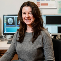
Webinar on Brain Imaging
Sprekers: Henk-(Jan) Mutsaerts & Nathalia Petridou
Recorded on march 17, 2022
Share this

Developing ASL perfusion MRI as cerebroVAScular biomarker of COgnitive Decline (VASCODE)decline
Henk(-Jan) Mutsaerts, MD PhD, Assistant professor, Department of Radiology & Nuclear Medicine, Amsterdam UMC
Short Abstract
This project aims to understand how changes in non-invasive brain perfusion MRI can predict cognitive decline in our aging society and its relationship with cardiovascular risk factors. While current imaging techniques can only identify irreversible late-stage structural damage, perfusion MRI is sensitive to early-stage cerebroVAScular changes that put patients at risk for COgnitive DEcline (VASCODE). To develop the full potential of perfusion MRI as a cerebrovascular biomarker, we: I) pool and harmonize multiple perfusion MRI from different scanners and populations to create the ASL-Based Brain Atlas (ABBA), II) disentangle on population-level different effects of normal physiological variability of perfusion MRI, and III) model associations between cardiovascular risk factors, brain perfusion, and cognitive decline. Our research may result in a non-invasive cerebrovascular biomarker that 1) increases our knowledge on the role of brain perfusion in cognitive decline, 2) monitors potential disease-modifying treatment effects, and eventually 3) helps predict cognitive decline.

From Blood to Neuron: High resolution functional imaging at 7 Tesla
Dr. Petridou pursued her doctoral research at the National Institutes of Health, Bethesda, MD, under the collaborative education program with the George Washington University, Washington DC, USA. After receiving her Doctor of Science (2006) she spent about three years as a Mansfield fellow at the Sir Peter Mansfield Magnetic Resonance Center, University of Nottingham, UK. In 2009 she joined the UMC Utrecht (Rudolf Magnus Institute and Radiology department), and is presently an Associate Professor with Radiology Dept. Dr. Petridou received a VIDI (STW) in 2013, for her research program on measuring neuronal activity in humans with high field fMRI.
Short Abstract
Functional magnetic resonance imaging (fMRI) measures brain activity via the accompanying changes in blood flow, volume, and oxygenation. As an indirect measure, the accuracy of fMRI is contingent on how closely the measured hemodynamic signals map onto the underlying neuronal activation patterns. The most specific hemodynamic signals arise from the capillary bed, the smallest vessels within gray matter (<20um) that directly serve active neuronal sites. At clinical field strengths, signals from larger veins at the surface of gray matter typically hinder the accuracy of fMRI. 7T allows for improved accuracy in two ways; first, signals from capillaries are enhanced while those from larger veins are reduced. Second, the high signal-to-noise allows for high spatial resolutions that can be exploited to pinpoint the vascular origin of signals measured according to physical location. This talk will give a view into how these advantages translate to increased fMRI specificity to neuronal activation patterns, in light of the correspondence of 7T BOLD fMRI to neuronal activity measured with intracranial electrophysiology (ECoG), in humans. An overview of how the improvements in specificity are being used to measure activity in small neuronal populations, at the spatial scale of columns and layers, will be presented.